Surgical imaging
Thanks to imec’s compact, video-rate on-chip spectral sensors, next-gen operating tools can offer surgeons an extra layer of crucial information in real time.
An extra pair of eyes in the operating theatre. What’s more, eyes with beyond-human vision. That’s the promise of spectral surgical imaging. It will guide surgeons to make better decisions, making their work easier and improving their patients’ chances.
Spectral surgical imaging can give surgeons a better view of:
- the precise borders of organs and tumors
- oxygenation of blood flowing through arteries and vessels
- biological, functional, and structural features of tissues
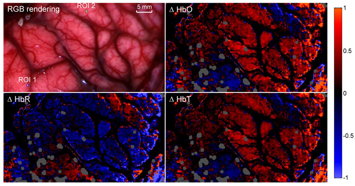
Snapshot spectral image of the human brain under guided surgery: the HSI camera is mounted onto a Carl ZEISS surgery microscope. – Courtesy of Polytechnique Montreal.
Image-guided surgery is sometimes achieved by injecting fluorescent contrast agents. Not ideal, because the number of approved dyes without side effects is limited. What’s more, this can be done only once during surgery – and only for a moment.
Other methods of surgical imaging – for instance based on CT and MRI scans– rely on highly specialized personnel and complex tools in the operating theatre. They disturb the operation flow and increase operating time. Underlining the need for additional surgical imaging methods that gives fast results, is non-invasive, and easy to use.
What spectral imaging can reveal
Spectral imaging is a non-invasive technique based on the principle that the light reflected, absorbed, ... by tissues in many bands of the electromagnetic spectrum (more than those discernable by the naked eye) reveals something about their biological make-up.
More about medical spectral imaging
Watch this video for a quick introduction to spectral imaging and imec’s technology:
There are many experimental use cases in medical literature that prove the value of spectral imaging for diagnostic and surgical applications. Examples are the discrimination of normal and tumor tissue during brain surgery, and the monitoring of blood flow and oxygen content during intestinal surgery.
Surgical spectral imaging needs compact, video-rate sensors
There’s plenty of evidence for the benefits of using spectral imaging in the operating room. So why are there barely any applications in clinical practice? One of the reasons is the difficulty of integrating spectral imaging in the surgical workflow.
For one thing, because we’re dealing with living tissue, a useful surgeon’s tool often needs to provide a moving image. Spectral systems that rely on spatial scanning are therefore not up to the task.
Secondly, most feasibility studies of surgical spectral imaging are done with bulky hardware that isn’t suitable for clinical practice. If we really want to integrate spectral cameras in (minimally invasive) operation flows, they’ll at least need to fit on standard laparoscopes.
Surgical spectral imaging from research to practice
By leveraging imec’s unique on-chip spectral imaging technology, the medical device industry can finally make this transition of surgical spectral imaging from research to practice.
Imec’s sensors are so compact they fit on a standard laparoscope, and those of the SNAPSHOT variety are able to register spectral video. Based on standard chip manufacturing processes, they’re also exceptionally affordable – especially when produced in middle to high volumes.
Want to get involved in bringing spectral imaging to the surgical imaging market? We’re ready to assist you by granting access to our technology, expertise and wide network of partners from across the medical device value chain.